
Translate this page into:
Neuroendocrine and sarcomatoid variants of carcinoma esophagus – Northeast Indian study

*Corresponding author: Srinivas Bannoth, Department of Surgical Oncology, Dr. B. Borooah Cancer Institute, Guwahati, Assam, India. srinivasbannoth@gmail.com
-
Received: ,
Accepted: ,
How to cite this article: Kalita D, Bannoth S, Singh P, Yadav J. Neuroendocrine and sarcomatoid variants of carcinoma esophagus – Northeast Indian study. Int J Mol Immuno Oncol 2020;5(2):82-5.
Abstract
Objective:
Variants of esophageal carcinoma such as sarcomatoid carcinoma and neuroendocrine carcinoma (NEC) of esophagus are rare presentations. This is a retrospective case series of 11 cases of carcinoma esophagus with atypical pathological presentation.
Materials and Methods:
The study period was from March 2016 to January 2019. Survival was estimated using Kaplan–Meier survival analysis method and comparison between groups made using log-rank test. P < 0.05 was considered as statistically significant at 95% confidence interval.
Results:
Eleven patients were included in the study, of which eight patients were of NEC of esophagus and three patients of sarcomatoid carcinoma of esophagus. The mean age of patients was 52.27 years. The overall survival at the end of the study was 0% as all patients in study were dead, overall median survival was 4.5 months. Patients who received palliative chemotherapy had median survival of 3 months, with palliative radiotherapy survival was 5 months. Treatment with chemotherapy + radiotherapy had median survival of 6 months and 2 months of median survival with supportive care.
Conclusion:
Esophageal NECs are rare and have aggressive presentation as seen in our study, all of our patients presented in advanced stage and multimodality treatment confers a better survival. Sarcomatoid carcinoma of esophagus usually presents at early stage and has better prognosis when compared to squamous cell carcinoma of esophagus, our patients presented at an advanced stage. Adequate pre-operative stabilization, multimodality treatment is a key for better outcome. Further large sample studies are needed to find out optimal treatment modalities.
Keywords
Esophageal carcinoma
Neuroendocrine carcinoma
Sarcomatoid carcinoma

INTRODUCTION
Variants of esophageal carcinoma such as sarcomatoid carcinoma and neuroendocrine carcinoma (NEC) of esophagus are rare presentations.[1-4] Sarcomatoid carcinoma of esophagus was first described by Virchow in 1865. On histopathology, they are characterized by biphasic components with bulk of tumor constituted with pleomorphic sarcomatoid appearance and few scattered areas of epithelial component.
Neuroendocrine neoplasms (NEN) of esophagus are rare which represent only 0.04–0.14% of gastrointestinal NEN.[1,2,5] NENs are epithelial neoplasms with predominant neuroendocrine differentiation.[1,3] They are categorized into morphological types, small cell and large cell types. About 90% of cases are small cell carcinoma (oat cell carcinoma).[6] The World Health Organization definition for NEC of esophagus includes tumors which are positive for chromogranin A, synaptophysin, and CD56 with a mitotic index of 20% or more.[7,8] Data from the US surveillance, epidemiology, and end results show that there is increasing trend in these tumors in view of improved diagnostic techniques.[9]
Prognosis of patients with sarcomatoid carcinoma tends to be better than squamous cell carcinoma as these tumors grow intraluminal and not in the wall. These tumors are typically confined to lamina propria and submucosa and rarely penetrate beyond submucosa.[10] Most of the neuroendocrine tumors present with lymph node or widespread metastasis at the time of initial diagnosis and are associated with poor prognosis in spite of multimodality treatment.[11-14]
MATERIALS AND METHODS
Patients diagnosed with neuroendocrine and sarcomatoid variants carcinoma of esophagus were taken into study. The study period was from March 2016 to November 2018. Diagnosis was made with upper gastrointestinal endoscopy and biopsy, which was later subjected to histopathology and immunohistochemistry [Figures 1 and 2]. Staging was done according to tumor, node, and metastasis (TNM) classification (8th edition of American Joint Committee on Cancer) for esophageal carcinoma. Data were collected from hospital records and telephonic conversation. Statistical Package for the Social Sciences version 16 software was used for analysis. Survival was estimated using Kaplan–Meier survival analysis method and comparison between groups was made using log-rank test. P < 0.05 was considered as statistically significant at 95% confidence interval.
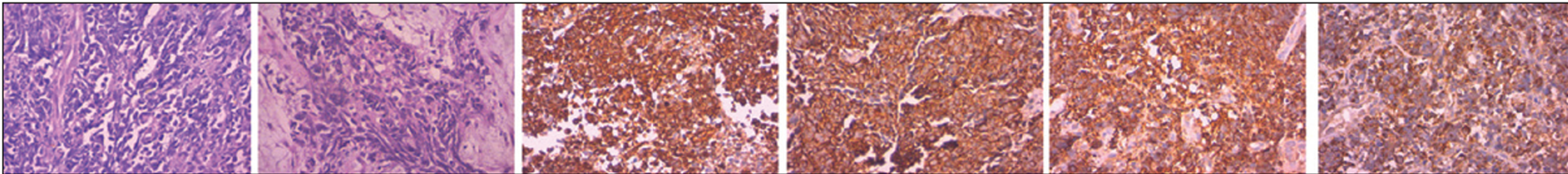
- Histopathology and immunohistochemistry pictures of neuroendocrine carcinoma.

- Histopathology and immunohistochemistry pictures of sarcomatoid carcinoma.
RESULTS
Eleven patients were included in the study, of which eight patients were of NEC of esophagus and three patients were of sarcomatoid carcinoma of esophagus. The mean age of patients was 52.27 years. Seven patients were male and four females. Most of the patients presented with ECOG performance score of 2 and 3 [Table 1].
| Clinical characteristics | Number |
|---|---|
| Mean age | 52.27 |
| Sex | |
| Male | 7 |
| Female | 4 |
| Pathology | |
| Neuroendocrine carcinoma | 8 |
| Sarcomatoid carcinoma | 3 |
| Level of esophagus | |
| Upper | 1 |
| Middle | 6 |
| Middle+lower | 3 |
| Lower | 1 |
| TNM stage | |
| IV A | 8 |
| IV B | 3 |
| Treatment received | |
| Palliative chemotherapy | 4 |
| Palliative radiotherapy | 2 |
| Chemotherapy+radiotherapy | 2 |
| Supportive care | 3 |
Mean hemoglobin of patients was 8.2 g/dl. Mean albumin of patients was 2.9 g/dl.
Involvement of upper cervical esophagus was seen in one patient, three patients had involvement of both middle and lower esophagus, and rest of them had involvement of either middle or lower part of esophagus. Out of 11 patients, eight of them presented with clinical TNM classification of clinical Stage IV A and rest of three patients presented with clinical Stage IV B [Table 1]. Lymph node metastasis was present in most of the patients. Distant metastasis in the form of liver and peritoneal metastasis was seen in three patients.
Palliative chemotherapy, palliative radiotherapy, chemotherapy+ radiotherapy, and best supportive care were given in four, two, two, and three patients, respectively [Table 1].
Kaplan–Meier survival curves showed that overall survival at the end of the study was 0% as all the patients in study were dead and overall median survival was 4.5 months [Figure 3]. Median survival of patients with NEC and sarcomatoid carcinoma was 2 and 6 months, respectively [Figure 4] with P = 0.120. Median survival according to the levels of esophagus involvement was 6 months for upper esophageal involvement, 2 months for middle esophageal level, 3 months for involvement of both middle and lower esophagus, and 5 months for lower esophageal involvement (P = 0.982) [Table 2]. Median survival for patients with Stage IV A was 4.5 months and that of Stage IV B was 3 months (P = 0.893).

- Kaplan–Meier curve for overall median survival.

- Kaplan–Meier curve for overall median survival for neuroendocrine and sarcomatoid carcinoma.
| Variable | Median survival (months) |
|---|---|
| Pathology | |
| Neuroendocrine carcinoma | 2 (P=0.120) |
| Sarcomatoid carcinoma | 6 (P=0.120) |
| Location | |
| Upper | 6 (P=0.982) |
| Middle | 2 (P=0.982) |
| Middle+lower | 3 (P=0.982) |
| Lower | 5 (P=0.982) |
| Stage | |
| IV A | 4.5 (P=0.893) |
| IV B | 3 (P=0.893) |
| ECOG score at presentation | |
| Score 2 | 6 (P=0.115) |
| Score 3 | 2 (P=0.115) |
| Treatment received | |
| Supportive care | 2 (P=0.19) |
| Palliative radiotherapy | 5 (P=0.19) |
| Palliative chemotherapy | 3 (P=0.19) |
| Chemotherapy+radiotherapy | 6 (P=0.19) |
Median survival of patients with palliative chemotherapy, palliative radiotherapy, chemotherapy+ radiotherapy, and supportive care was 3, 5, 6, and 2 months, respectively (P = 0.19). Median survival of patients with performance status ECOG-2 was 6 months and with of ECOG-3 was 2 months with P = 0.115 [Table 2].
DISCUSSION
Adenocarcinoma and squamous cell carcinomas are two unique entities of esophageal carcinoma. Variants of esophageal carcinoma such as sarcomatoid carcinoma and NEC of esophagus are rare presentations.[1-4] NENs of esophagus are rare which represent only 0.04–0.14% of gastrointestinal NEN.[1,2,5] They are categorized into morphological types, small cell and large cell type. About 90% of cases are small cell carcinoma type also called as oat cell carcinoma.[6] Sarcomatoid carcinomas usually represent 2% of esophageal carcinomas.
Esophageal NEC and sarcomatoid carcinoma usually predominate in males.[16] Demographic data of our study showed male:female ratio of 1.75:1. NECs are usually solitary lesions and develop in the lower third of esophagus.[7,12,15] In our study, involvement was seen in middle and lower part of esophagus. Sarcomatoid carcinoma of esophagus presents as polypoidal lesions that may reach a diameter of up to 15 cm. Surface of lesions is smooth and tumors are attached by a pedicle, occasionally, there is no pedicle. The depth of invasion determines the risk of metastasis (10% for involvement of lamina propria, 25% for tumors reaching submucosa, and 75% for tumors, which penetrate adventitia).
Most of our patients presented with poor performance status and in advanced stage with severe malnutrition. After discussion in institutional multidisciplinary tumor board, palliative chemotherapy was given in four patients, two patients had palliative radiotherapy, two patients had chemotherapy along with radiotherapy, and three patients were given only palliative supportive care. None of our patients underwent surgery in view of poor general condition and advanced stage of disease. This represents aggressive nature and poor prognosis of NEC of esophagus. Although sarcomatoid carcinoma usually does not penetrate beyond submucosa, our patients had late presentation with advanced disease. Overall survival at the end of the study was 0% and median survival of 4.5 months, as shown in Figure 3.
Patients who presented with good performance status were treated with chemotherapy+ radiotherapy and they had relative better survival when compared to palliative therapies. Patients who received palliative radiotherapy had better survival than palliative chemotherapy. Survival of patient who was treated only with best supportive care was least. The risk of death was 2.78 times higher in patients with ECOG-3 than ECOG-2 status [Table 2].
Limitation of our study was small sample size, none of patient underwent surgical treatment to evaluate outcomes of surgery and all the patients were of Stage IV so outcomes of patients with early stages could not be studied.
CONCLUSION
Esophageal NECs are rare and have aggressive presentation as seen in our study, all of our patients presented in advanced stage. Sarcomatoid carcinoma of esophagus usually presents at early stage and has better prognosis when compared to squamous cell carcinoma of esophagus, but our patients presented at an advanced stage. Adequate pre-operative stabilization, multimodality treatment may lead to better outcome. Further large sample studies are needed to find out optimal treatment modalities of these entities.
Acknowledgments
Our patients, Department of Surgical oncology.
Declaration of patient consent
Patient’s consent not required as patients identity is not disclosed or compromised.
Financial support and sponsorship
Nil.
Conflicts of interest
There are no conflicts of interest.
References
- The clinical features and treatment modality of esophageal neuroendocrine tumors: A multicenter study in Korea. BMC Cancer. 2014;14:569.
- [CrossRef] [PubMed] [Google Scholar]
- Primary small cell carcinoma of the esophagus: Clinicopathological and immunohistochemical features of 21 cases. BMC Cancer. 2007;7:38.
- [CrossRef] [PubMed] [Google Scholar]
- Neuroendocrine tumors involving the gastroenteropancreatic tract: A clinicopathological evaluation of 773 cases. Clinics (Sao Paulo). 2011;66:1671-5.
- [Google Scholar]
- Variants of squamous cell carcinoma In: Surgical Pathology of the GI Tract, Liver, Biliary Tract and Pancreas (2nd ed). Philadelphia, PA: Saunders Elsevier; 2009. p. :548-52.
- [Google Scholar]
- Current trends of the incidence and pathological diagnosis of gastroenteropancreatic neuroendocrine tumors (GEP-NETs) in Korea 2000-2009: Multicenter study. Cancer Res Treat. 2012;44:157-65.
- [Google Scholar]
- Oat-cell carcinoma of the oesophagus. J Pathol Bacteriol. 1952;64:889-91.
- [CrossRef] [PubMed] [Google Scholar]
- Primary high-grade neuroendocrine carcinoma of the esophagus: A clinicopathologic and immunohistochemical study of 42 resection cases. Am J Surg Pathol. 2013;37:467-83.
- [CrossRef] [PubMed] [Google Scholar]
- Neuroendocrine Neoplasms of the Oesophagus. Lyons, France: IARC Press; 2010.
- An analysis of rare carcinoid tumors: Clarifying these clinical conundrums. World J Surg. 2005;29:92-101.
- [CrossRef] [PubMed] [Google Scholar]
- Gastroenteropancreatic neuroendocrine tumours. Lancet Oncol. 2008;9:61-72.
- [CrossRef] [Google Scholar]
- Special variants of squamous cell carcinoma In: Lewin KJ, Appelman HD, eds. Atlas of Tumor Pathology, Third series, Fascicle 18: Tumors of the Esophagus and Stomach. Washington, DC: Armed Forces Institute of Pathology; 1996. p. :83-91.
- [Google Scholar]
- Small cell carcinoma of the esophagus: Analysis of 10 cases and review of the published data. Am J Clin Oncol. 2000;23:455-9.
- [CrossRef] [PubMed] [Google Scholar]
- Primary undifferentiated small cell carcinoma of the esophagus. Hum Pathol. 1999;30:216-21.
- [CrossRef] [Google Scholar]
- Retrospective study of clinicopathologic features and prognosis of high-grade neuroendocrine carcinoma of the esophagus. Am J Surg Pathol. 2008;32:1404-11.
- [CrossRef] [PubMed] [Google Scholar]
- Small cell carcinoma of the esophagus. Report of three cases and review of the literature. Dig Dis Sci. 1990;35:145-52.
- [CrossRef] [PubMed] [Google Scholar]
- Neuroendocrine tumor of the esophagus-successful endoscopic treatment of a very rare entity. Dtsch Med Wochenschr. 2010;135:19-21.
- [CrossRef] [PubMed] [Google Scholar]


